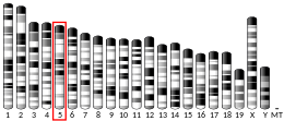
PPARGC1A
PPARGC1AまたはPGC-1α(peroxisome proliferator-activated receptor gamma coactivator 1-alpha)は、ヒトではPPARGC1A遺伝子にコードされるタンパク質である[4]。PPARGC1Aはhuman accelerated regionと呼ばれる、チンパンジーとの共通祖先からの分岐以降に塩基置換率が加速しているゲノム領域(HAR20)と関係しており、そのため類人猿からヒトの分岐に重要な役割を果たした可能性がある[5]。
PGC-1αは、ミトコンドリア生合成のマスターレギュレーターである[6][7][8]。また、PGC-1αは肝臓における糖新生の主要な調節因子であり、糖新生のための遺伝子発現の増加などを担う[9]。
機能[編集]
PGC-1αはエネルギー代謝に関与する遺伝子を調節する転写コアクチベーターであり、ミトコンドリア生合成のマスターレギュレーターである[6][7][8]。このタンパク質は核内受容体のPPARγと相互作用し、それによって複数の転写因子との相互作用が可能となる。また、このタンパク質はCREBや核呼吸因子(nuclear respiratory factor, NRF)とも相互作用し、これらの活性を調節する[10]。PGC-1αは外部の生理的刺激とミトコンドリア生合成の調節を直接関連付ける役割を果たし[10]、また筋繊維のタイプの分化を調節する主要な因子でもあり、遅筋繊維の形成を駆動する[11]。
持久運動はヒトの骨格筋においてPGC-1αの遺伝子を活性化することが示されている[12]。運動によって骨格筋で誘導されたPGC-1αはオートファジー[13]と小胞体ストレス応答[14]を増大させる。
PGC-1αタンパク質は、血圧の制御、細胞のコレステロール恒常性の調節、そして肥満に関与している可能性がある[10]。
PGC-1αによるSIRT3のアップレギュレーションは、ミトコンドリアをより健全にする[15]。
調節[編集]
PGC-1αは外部からのシグナルを統合する主要な因子であると考えられている。PGC-1αは多くの因子によって活性化されることが知られている。
- 絶食は肝臓のPGC-1αなど、糖新生に関与する遺伝子の発現を増加させる[16][17]。
- 低温曝露によって強く誘導され、この環境刺激を適応的熱産生(adaptive thermogenesis)へ関連付ける[18]。
- 持久運動によって誘導される[12]。PGC-1αは乳酸代謝を決定する。持久運動時の乳酸の高レベルの蓄積を防ぎ、乳酸をエネルギー源としてより効率的に利用できるようにする[19]。
- SIRT1はPGC-1αに結合して脱アセチル化によって活性化し、ミトコンドリア生合成に影響を与えることなく糖新生を誘導する[20]。
PGC-1αは上流の調節因子の一部に対してポジティブフィードバックを行うことが示されている。
AktとカルシニューリンはどちらもNF-κB(p65)の活性化因子である[23][24]。PGC-1αはこれらを活性化することでNF-κBを活性化しているようである。筋肉ではPGC-1αの誘導後にNF-κB活性の増大がみられることが示されているが[25]、この発見には議論があり、他のグループはPGC-1がNF-κBの活性を阻害することを示している[26]。
PGC-1αは急性腎障害時にNADの生合成を駆動し、腎臓の保護に大きな役割を果たすことが示されている[27]。
臨床的意義[編集]
PPARGGC1Aはミトコンドリア代謝に対する保護効果によってパーキンソン病の治療となる可能性が示唆されている[28]。
さらに、PGC-1αの脳特異的アイソフォームはハンチントン病、筋萎縮性側索硬化症など他の神経変性疾患に役割を果たしている可能性が高いことが同定されている[29][30]。
マッサージ治療はPGC-1αの量を増加させ、新たなミトコンドリアの産生をもたらすようである[31][32][33]。
PGC-1αとβは、STAT6の上流の活性化に伴うPPARγとの相互作用によって、抗炎症性M2マクロファージの極性化に関与することが示唆されている。PGC-1のSTAT6/PPARγを介したM2マクロファージ活性化効果は独立した研究でも確認されており、さらにPGC-1が炎症性サイトカインの産生を阻害することも示されている[34]。
PGC-1αは運動中の筋肉から3-アミノイソ酪酸の分泌を担うことが提唱されている[35]。白色脂肪における3-アミノイソ酪酸の効果には、白色脂肪組織の褐色化を促進する熱産生遺伝子の活性化や、その後のバックグラウンド代謝の増加などがある。したがって、3-アミノイソ酪酸はPGC-1αのメッセンジャー分子として作用し、白色脂肪など他の組織でPGC-1α増大の効果がみられることが説明される。
相互作用[編集]
PPARGC1Aは次に挙げる因子と相互作用することが示されている。
- CREB結合タンパク質[36]
- エストロゲン関連受容体α(ERRα)[37]、エストロゲン関連受容体β(ERRβ)、エストロゲン関連受容体γ(ERRγ)
- ファルネソイドX受容体[38]
- FBXW7[39]
- MED1[40]、MED12[40]、MED14[40]、MED17[40]
- NRF1[41]
- PPARγ[36][40]
- レチノイドX受容体α[42]
- 甲状腺ホルモン受容体β[43]
出典[編集]
- ^ a b c GRCm38: Ensembl release 89: ENSMUSG00000029167 - Ensembl, May 2017
- ^ Human PubMed Reference:
- ^ Mouse PubMed Reference:
- ^ “Human peroxisome proliferator activated receptor gamma coactivator 1 (PPARGC1) gene: cDNA sequence, genomic organization, chromosomal localization, and tissue expression”. Genomics 62 (1): 98–102. (Feb 2000). doi:10.1006/geno.1999.5977. PMID 10585775.
- ^ “An RNA gene expressed during cortical development evolved rapidly in humans”. Nature 443 (7108): 167–72. (September 2006). Bibcode: 2006Natur.443..167P. doi:10.1038/nature05113. PMID 16915236.
- ^ a b “Mitochondrial biogenesis: pharmacological approaches”. Curr. Pharm. Des. 20 (35): 5507–9. (2014). doi:10.2174/138161282035140911142118. hdl:10454/13341. PMID 24606795. "Mitochondrial biogenesis is therefore defined as the process via which cells increase their individual mitochondrial mass [3]. ... This work reviews different strategies to enhance mitochondrial bioenergetics in order to ameliorate the neurodegenerative process, with an emphasis on clinical trials reports that indicate their potential. Among them creatine, Coenzyme Q10 and mitochondrial targeted antioxidants/peptides are reported to have the most remarkable effects in clinical trials."
- ^ a b “Mitochondrial biogenesis in health and disease. Molecular and therapeutic approaches”. Curr. Pharm. Des. 20 (35): 5619–5633. (2014). doi:10.2174/1381612820666140306095106. PMID 24606801. "Mitochondrial biogenesis (MB) is the essential mechanism by which cells control the number of mitochondria."
- ^ a b “Mitochondrial biogenesis and dynamics in the developing and diseased heart”. Genes Dev. 29 (19): 1981–91. (2015). doi:10.1101/gad.269894.115. PMC 4604339. PMID 26443844.
- ^ “Biological and catalytic functions of sirtuin 6 as targets for small-molecule modulators”. Journal of Biological Chemistry 295 (32): 11021–11041. (2020). doi:10.1074/jbc.REV120.011438. PMC 7415977. PMID 32518153.
- ^ a b c “PPARGC1A PPARG coactivator 1 alpha [Homo sapiens (human) - Gene - NCBI]”. www.ncbi.nlm.nih.gov. 2022年3月5日閲覧。
- ^ “Transcriptional co-activator PGC-1 alpha drives the formation of slow-twitch muscle fibres”. Nature 418 (6899): 797–801. (2002). Bibcode: 2002Natur.418..797L. doi:10.1038/nature00904. PMID 12181572.
- ^ a b “Exercise induces transient transcriptional activation of the PGC-1alpha gene in human skeletal muscle”. J. Physiol. 546 (Pt 3): 851–8. (February 2003). doi:10.1113/jphysiol.2002.034850. PMC 2342594. PMID 12563009.
- ^ “Role of PGC-1α during acute exercise-induced autophagy and mitophagy in skeletal muscle”. American Journal of Physiology 308 (9): C710-719. (2015). doi:10.1152/ajpcell.00380.2014. PMC 4420796. PMID 25673772.
- ^ “The unfolded protein response mediates adaptation to exercise in skeletal muscle through a PGC-1α/ATF6α complex”. Cell Metabolism 13 (2): 160–169. (2011). doi:10.1016/j.cmet.2011.01.003. PMC 3057411. PMID 21284983.
- ^ “Mitochondrial dysregulation and muscle disuse atrophyy”. F1000Research 8: 1621. (2019). doi:10.12688/f1000research.19139.1. PMC 6743252. PMID 31559011.
- ^ Canettieri, G., Koo, S.-H., Berdeaux, R., Heredia, J., Hedrick, S., Zhang, X., & Montminy (2005). “Dual role of the coactivator TORC2 in modulating hepatic glucose output and insulin signaling”. Cell Metabolism 2 (5): 331–338. doi:10.1016/j.cmet.2005.09.008. PMID 16271533.
- ^ Yoon, J. Cliff; Puigserver, Pere; Chen, Guoxun; Donovan, Jerry; Wu, Zhidan; Rhee, James; Adelmant, Guillaume; Stafford, John et al. (September 2001). “Control of hepatic gluconeogenesis through the transcriptional coactivator PGC-1” (英語). Nature 413 (6852): 131–138. Bibcode: 2001Natur.413..131Y. doi:10.1038/35093050. ISSN 1476-4687. PMID 11557972.
- ^ “PGC-1alpha: a key regulator of energy metabolism”. Adv Physiol Educ 30 (4): 145–51. (December 2006). doi:10.1152/advan.00052.2006. PMID 17108241.
- ^ “Skeletal muscle PGC-1α controls whole-body lactate homeostasis through estrogen-related receptor α-dependent activation of LDH B and repression of LDH A”. Proc. Natl. Acad. Sci. U.S.A. 110 (21): 8738–43. (May 2013). Bibcode: 2013PNAS..110.8738S. doi:10.1073/pnas.1212976110. PMC 3666691. PMID 23650363.
- ^ “Nutrient control of glucose homeostasis through a complex of PGC-1alpha and SIRT1”. Nature 434 (7029): 113–8. (March 2005). Bibcode: 2005Natur.434..113R. doi:10.1038/nature03354. PMID 15744310.
- ^ “Myopathy caused by mammalian target of rapamycin complex 1 (mTORC1) inactivation is not reversed by restoring mitochondrial function”. Proc. Natl. Acad. Sci. U.S.A. 108 (51): 20808–13. (December 2011). Bibcode: 2011PNAS..10820808R. doi:10.1073/pnas.1111448109. PMC 3251091. PMID 22143799.
- ^ “Remodeling of calcium handling in skeletal muscle through PGC-1α: impact on force, fatigability, and fiber type”. Am. J. Physiol., Cell Physiol. 302 (1): C88–99. (January 2012). doi:10.1152/ajpcell.00190.2011. PMID 21918181.
- ^ “Phosphorylation of NF-kappaB and IkappaB proteins: implications in cancer and inflammation”. Trends Biochem. Sci. 30 (1): 43–52. (January 2005). doi:10.1016/j.tibs.2004.11.009. PMID 15653325.
- ^ “Multiple roles of the DSCR1 (Adapt78 or RCAN1) gene and its protein product calcipressin 1 (or RCAN1) in disease”. Cell. Mol. Life Sci. 62 (21): 2477–86. (November 2005). doi:10.1007/s00018-005-5085-4. PMID 16231093.
- ^ “Skeletal muscle PGC-1α is required for maintaining an acute LPS-induced TNFα response”. PLOS ONE 7 (2): e32222. (2012). Bibcode: 2012PLoSO...732222O. doi:10.1371/journal.pone.0032222. PMC 3288087. PMID 22384185.
- ^ “Peroxisome proliferator-activated receptor gamma coactivator 1alpha or 1beta overexpression inhibits muscle protein degradation, induction of ubiquitin ligases, and disuse atrophy”. J. Biol. Chem. 285 (25): 19460–71. (June 2010). doi:10.1074/jbc.M110.113092. PMC 2885225. PMID 20404331.
- ^ “PGC1α drives NAD biosynthesis linking oxidative metabolism to renal protection”. Nature 531 (7595): 528–32. (2016). Bibcode: 2016Natur.531..528T. doi:10.1038/nature17184. PMC 4909121. PMID 26982719.
- ^ “PGC-1{alpha}, A Potential Therapeutic Target for Early Intervention in Parkinson's Disease”. Sci Transl Med 2 (52): 52ra73. (October 2010). doi:10.1126/scitranslmed.3001059. PMC 3129986. PMID 20926834.
- ^ “A greatly extended PPARGC1A genomic locus encodes several new brain-specific isoforms and influences Huntington disease age of onset”. Human Molecular Genetics 21 (15): 3461–73. (2012). doi:10.1093/hmg/dds177. PMID 22589246.
- ^ “PGC-1α is a male-specific disease modifier of human and experimental amyotrophic lateral sclerosis”. Human Molecular Genetics 22 (17): 3477–84. (2013). doi:10.1093/hmg/ddt202. PMID 23669350.
- ^ “Massage therapy attenuates inflammatory signaling after exercise-induced muscle damage”. Sci Transl Med 4 (119): 119ra13. (February 2012). doi:10.1126/scitranslmed.3002882. PMID 22301554.
- ^ “Study works out kinks in understanding of massage”. Los Angeles Times. (2012年2月1日)
- ^ “Videos | The Buck Institute for Research on Aging”. Buckinstitute.org. 2013年10月11日閲覧。
- ^ “Oxidative metabolism and PGC-1beta attenuate macrophage-mediated inflammation”. Cell Metab. 4 (1): 13–24. (July 2006). doi:10.1016/j.cmet.2006.05.011. PMC 1904486. PMID 16814729.
- ^ “β-Aminoisobutyric acid induces browning of white fat and hepatic β-oxidation and is inversely correlated with cardiometabolic risk factors”. Cell Metabolism 19 (1): 96–108. (2014). doi:10.1016/j.cmet.2013.12.003. PMC 4017355. PMID 24411942.
- ^ a b “Activation of PPARgamma coactivator-1 through transcription factor docking”. Science 286 (5443): 1368–71. (November 1999). doi:10.1126/science.286.5443.1368. PMID 10558993.
- ^ “The estrogen-related receptor alpha (ERRalpha) functions in PPARgamma coactivator 1alpha (PGC-1alpha)-induced mitochondrial biogenesis”. Proceedings of the National Academy of Sciences of the United States of America 101 (17): 6472–7. (April 2004). Bibcode: 2004PNAS..101.6472S. doi:10.1073/pnas.0308686101. PMC 404069. PMID 15087503.
- ^ “Peroxisome proliferator-activated receptor-γ coactivator 1α (PGC-1α) regulates triglyceride metabolism by activation of the nuclear receptor FXR”. Genes Dev. 18 (2): 157–69. (January 2004). doi:10.1101/gad.1138104. PMC 324422. PMID 14729567.
- ^ “SCFCdc4 acts antagonistically to the PGC-1α transcriptional coactivator by targeting it for ubiquitin-mediated proteolysis”. Genes Dev. 22 (2): 252–64. (January 2008). doi:10.1101/gad.1624208. PMC 2192758. PMID 18198341.
- ^ a b c d e “Coordination of p300-mediated chromatin remodeling and TRAP/mediator function through coactivator PGC-1alpha”. Mol. Cell 12 (5): 1137–49. (November 2003). doi:10.1016/S1097-2765(03)00391-5. PMID 14636573.
- ^ “Mechanisms controlling mitochondrial biogenesis and respiration through the thermogenic coactivator PGC-1”. Cell 98 (1): 115–24. (1999). doi:10.1016/S0092-8674(00)80611-X. PMID 10412986.
- ^ “PGC-1 functions as a transcriptional coactivator for the retinoid X receptors”. J. Biol. Chem. 277 (6): 3913–7. (February 2002). doi:10.1074/jbc.M109409200. PMID 11714715.
- ^ “Requirement of helix 1 and the AF-2 domain of the thyroid hormone receptor for coactivation by PGC-1”. J. Biol. Chem. 277 (11): 8898–905. (March 2002). doi:10.1074/jbc.M110761200. PMID 11751919.
関連文献[編集]
- “PGC-1, a versatile coactivator”. Trends Endocrinol. Metab. 12 (8): 360–5. (2001). doi:10.1016/S1043-2760(01)00457-X. PMID 11551810.
- “Peroxisome proliferator-activated receptor-gamma coactivator 1 alpha (PGC-1 alpha): transcriptional coactivator and metabolic regulator”. Endocr. Rev. 24 (1): 78–90. (2003). doi:10.1210/er.2002-0012. PMID 12588810.
- “PGC-1alpha: a potent transcriptional cofactor involved in the pathogenesis of type 2 diabetes”. Diabetologia 49 (7): 1477–88. (2007). doi:10.1007/s00125-006-0268-6. PMID 16752166.
- “Peroxisome proliferator-activated receptor gamma coactivator 1 coactivators, energy homeostasis, and metabolism”. Endocr. Rev. 27 (7): 728–35. (2007). doi:10.1210/er.2006-0037. PMID 17018837.
関連項目[編集]
外部リンク[編集]
- PPARGC1A protein, human - MeSH・アメリカ国立医学図書館・生命科学用語シソーラス(英語)
- FactorBook PGC1A
- Overview of all the structural information available in the PDB for UniProt: Q9UBK2 (Peroxisome proliferator-activated receptor gamma coactivator 1-alpha) at the PDBe-KB.


