「プロテオパチー」の版間の差分
ページ「Proteopathy」の翻訳により作成 |
(相違点なし)
|
2020年7月22日 (水) 11:00時点における版
| プロテオパチー | |
|---|---|
 | |
| 老人斑(senile plaques)と脳アミロイドアンギオパチー(脳アミロイド血管症)に蓄積するタンパク質断片である「アミロイドβ」(Aβ)(茶色)に対する抗体で免疫染色した、アルツハイマー病患者からの大脳皮質の切片の顕微鏡写真。10倍顕微鏡対物レンズ。 | |
| 概要 | |
| 分類および外部参照情報 |
医学では、プロテオパチー(proteopathy; プロテオパシーとも) ([proʊtiːˈɒpəθiː]; Proteo- [pref. protein]; -pathy [suff. disease]; proteopathies pl.; proteopathic adj)は、特定のタンパク質が構造的に異常になり、体の細胞、組織、臓器の機能を破壊する疾患のクラスを指す[1][2]。 多くの場合、タンパク質は正常な構成にフォールディング(折り畳み)できない。 このミスフォールディング(誤った折り畳み)状態では、タンパク質は何らかの方法で毒性になるか(毒性機能の獲得)、または通常の機能を失う可能性がある[3]。
プロテオパチー(proteopathies) (別名: プロテイノパチー(proteinopathies)、タンパク質構造障害(protein conformational disorders)、またはタンパク質のミスフォールディング疾患(protein misfolding diseases)として知られている)は、クロイツフェルト・ヤコブ病や他のプリオン病、アルツハイマー病、パーキンソン病、アミロイドーシス、多系統萎縮症、および他の疾患の広い範囲が含まれている(プロテオパチーのリストを参照)[2][4][5][6][7][8]。 プロテオパチー(proteopathy)という用語は、最初にラリーウォーカー(Lary Walker)とハリー・レヴィン(Harry LeVine)によって2000年に提案された[9]。
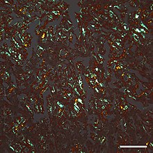
プロテオパシーの概念は、その起源を19世紀半ばまで遡ることができ、1854年、ルドルフ・ルートヴィヒ・カール・フィルヒョウ(Rudolf Virchow)は、アミロイド(amyloid)(「デンプンのような」)という用語を作り、セルロースに似た化学反応を示す大脳アミロイド小体の物質を説明した。1859年、フリードライヒ(Friedreich)とケクレ(Kekulé)は、「アミロイド」がセルロースからなるのではなく、実際にはタンパク質を豊富に含んでいることを示した[10]。 その後の研究では、多くの異なるタンパク質がアミロイドを形成し、すべてのアミロイドは、コンゴーレッド色素で染色した後の交差偏光での複屈折だけでなく、電子顕微鏡で見たときにフィブリル状(fibrillar; 繊維状)の超微細構造を持つという共通点を持っていることが示されている[10]。 しかし、いくつかの蛋白質性病変は複屈折を欠き、アルツハイマー病患者の脳内にAβタンパク質がびまん性に沈着しているような古典的なアミロイド線維をほとんど含まないか、または全く含まない[11]。 さらに、オリゴマーとして知られている小さな非フィブリル性タンパク質の集合体は罹患臓器の細胞に毒性があり、そのフィブリル性形態のアミロイド原性タンパク質は比較的良性である可能性があるという証拠が明らかになった[12][13]。

病態生理
ほとんどの場合、すべてのプロテオパチーではないにしても、3次元フォールディング(コンフォメーション)の変化により、特定のタンパク質がそれ自体に結合する傾向が高まる[14]。 この凝集形態では、タンパク質は除去(clearance; クリアランス)に対する抵抗性があり、影響を受ける臓器の正常な能力を妨害する可能性がある。 場合によっては、タンパク質のミスフォールディングにより、通常の機能が失われる。 例えば、嚢胞性線維症は、嚢胞性線維症膜貫通調節因子(CFTR)タンパク質の欠陥によって引き起こされ[15]、筋萎縮性側索硬化症/前頭側頭葉変性症(FTLD)では、特定の遺伝子調節タンパク質が細胞質内で不適切に凝集し、核内での通常の役割を実行できない[16][17]。 タンパク質は、ポリペプチド骨格として知られる共通の構造的特徴を共有しているため、すべてのタンパク質は、ある状況下でミスフォールドされる可能性がある[18]。 しかし、おそらく脆弱なタンパク質の構造的特異性のために、比較的少数のタンパク質のみがタンパク質変成疾患に関連している。 例えば、通常はアンフォールド(折り畳まれていない)されているか、またはモノマーとして比較的不安定なタンパク質(つまり、単一の非結合タンパク質分子)は、異常なコンフォメーションにミスフォールドする可能性が高くなる[14][18][19]。 ほぼ全ての場合において、疾患を引き起こす分子構成には、タンパク質のβシート二次構造の増加を伴う[14][18][20][21]。 いくつかのプロテオパチーにおける異常なタンパク質は、複数の3次元形状に折りたたまれることが示されてる。 これらの変性タンパク質構造は、それらの異なる病原性、生化学的、およびコンフォメーション特性によって定義される[22]。 これらはプリオン病に関して最も徹底的に研究されており、タンパク質株と呼ばれている[23][24]。

プロテオパシーが発症する可能性は、タンパク質の自己組織化を促進する特定の危険因子によって増加する。 これらは、タンパク質の一次アミノ酸配列の不安定化変化、翻訳後修飾(過剰リン酸化など)、温度やpHの変化、タンパク質の生産量の増加、またはその除去(クリアランス)の減少が含まれている[25][26][27]。 加齢は、外傷性脳損傷と同様に[28][29]、強い危険因子である[25]。 老化した脳では、複数のプロテオパチーが重複する可能性がある[30]。 例えば、タウオパチーとAβアミロイドーシス(アルツハイマー病の重要な病理学的特徴として共存する)に加えて、多くのアルツハイマー病患者は脳内にシヌクレイノパチー(レビー小体)を併発している[31]。
シャペロンやコ・シャペロン(タンパク質のフォールディングを助けるタンパク質)が、加齢や蛋白質ミスフォールディング病において、タンパク質の毒性に拮抗し、タンパク質恒常性を維持しているのではないかという仮説が立てられている[32][33][34]。
播種誘発(seeded induction)
いくつかのタンパク質は、疾患を引き起こすコンフォメーションに折り畳まれた同じ(または類似の)タンパク質への曝露によって、異常な集合体を形成するように誘導でき、これは「播種(seeding)」または「許容テンプレート化(permissive templating)」と呼ばれるプロセスである[35][36]。 このようにして、罹患したドナーから罹患組織抽出物を導入することにより、罹患性宿主に疾患状態を引き起こすことができる。 そのような誘導性プロテオパシーの最もよく知られている形態はプリオン病であり[37]、これは、疾患を引き起こすコンフォメーションの精製プリオンタンパク質に、宿主生物を曝露することによって感染する可能性がある[38][39]。 現在、Aβアミロイドーシス、アミロイドA(AA)アミロイドーシス、およびアポリポプロテインA-IIアミロイドーシス[36][40]、タウオパチー[41]、シヌクレイノパチー[42][43][44][45]、およびスーパーオキシドジスムターゼ-1(SOD1)[46][47]、ポリグルタミン[48][49]、およびTAR DNA結合タンパク-43(TDP-43)の凝集を含む[50]、他のプロテオパチーが同様のメカニズムによって誘発されるという証拠がある。
これらの例のすべてにおいて、タンパク質の異常な形態自体が病原体であるように見える。 場合によっては、あるタイプのタンパク質の沈着は、おそらくタンパク質分子の構造的相補性のために、βシート構造に富む他のタンパク質の集合体によって実験的に誘発されることがある。 例えば、AAアミロイドーシスは、絹、酵母アミロイドSup35、大腸菌(Escherichia coli)由来のカーリー線維(curli fibrils)などの多様な高分子によってマウスで刺激される[51]。 さらに、アポリポプロテインA-IIアミロイドは、βシートを豊富に含む様々なアミロイド原線維によってマウスで誘発され[52]、脳タウオパチーは、凝集したAβを豊富に含む脳抽出物によって誘導される[53]。 また、プリオンタンパク質とAβとの交雑播種(cross-seeding)の実験的証拠もある[54]。 一般に、このような異種播種は、同じタンパク質の破損した形態による播種よりも効率が悪い。
プロテオパシーのリスト
治療
多くのプロテオパチーのための効果的な治療法の開発は、挑戦的である[55][56]。 プロテオパチーは、多くの場合、異なる原因から生じる異なるタンパク質が関与しているため、治療戦略はそれぞれの疾患に合わせてカスタマイズする必要がある。 しかし、一般的な治療法としては、羅漢した臓器の機能を維持し、疾患の原因となるタンパク質の形成を減少させ、タンパク質のミスフォールディングおよび/または凝集を防止し、またはそれらの除去の促進が含まれる[57][55][58]。 例えば、アルツハイマー病では、疾患関連タンパク質Aβを親タンパク質から遊離させる酵素を阻害することにより、疾患関連タンパク質Aβの産生を減らす方法が研究されている[56]。 別の戦略は、抗体を用いて能動的または受動的な免疫化によって特定のタンパク質を中和することである[59]。 いくつかのプロテオパチーでは、タンパク質オリゴマーの毒性作用を阻害することが有益な場合がある[60]。 アミロイドA(AA)アミロイドーシスは、血中のタンパク質(血清アミロイドA、またはSAA呼ばれる)の量を増加させる炎症状態を治療することによって減少できる[55]。 免疫グロブリン軽鎖アミロイドーシス(ALアミロイドーシス)では、化学療法により、様々な体の臓器でアミロイドを形成する軽鎖タンパク質を作る血球の数を減らすことができる[61]。 トランスサイレチン(TTR)アミロイドーシス(ATTR)は、ミスフォールドされたTTRが複数の臓器に沈着することに起因する[62]。 TTRは主に肝臓で産生されるため、TTRアミロイドーシスは、一部の遺伝性症例では、肝移植により進行を遅らせられる可能性がある[63]。 TTRアミロイドーシスはまた、タンパク質の正常な集合体(4つのTTR分子が結合して構成されているため、テトラマーと呼ばれる)を安定化させることによって治療できる。 安定化により、個々のTTR分子が逃げたり、ミスフォールディングしたり、アミロイドに凝集するのを防ぐことができる[64][65]。
プロテオパチーのための他のいくつかの治療戦略が研究されているが、これには、低分子(small molecule)および低分子干渉RNA、アンチセンスオリゴヌクレオチド、ペプチド、および人工免疫細胞などの生物学的医薬品が含まれる[66][67][68][69]。 場合によっては、複数の治療薬を組み合わせて効果を高めることもある[67][70]。
追加画像
-
アルツハイマー病患者の大脳皮質の神経細胞体(矢印)と突起(process)(矢尻)のタウオパチー(茶色)の顕微鏡写真。バー=25ミクロン(0.025mm)。
参照
参考文献
- ^ “The cerebral proteopathies”. Neurobiology of Aging 21 (4): 559–61. (2000). doi:10.1016/S0197-4580(00)00160-3. PMID 10924770.
- ^ a b “The cerebral proteopathies: neurodegenerative disorders of protein conformation and assembly”. Molecular Neurobiology 21 (1–2): 83–95. (2000). doi:10.1385/MN:21:1-2:083. PMID 11327151.
- ^ “Protein misfolding and disease: from the test tube to the organism”. Current Opinion in Chemical Biology 12 (1): 25–31. (February 2008). doi:10.1016/j.cbpa.2008.02.011. PMID 18295611.
- ^ “Protein misfolding, functional amyloid, and human disease”. Annual Review of Biochemistry 75 (1): 333–66. (2006). doi:10.1146/annurev.biochem.75.101304.123901. PMID 16756495.
- ^ “Conformational disease”. Lancet 350 (9071): 134–8. (July 1997). doi:10.1016/S0140-6736(97)02073-4. PMID 9228977.
- ^ “A primer of amyloid nomenclature”. Amyloid 14 (3): 179–83. (September 2007). doi:10.1080/13506120701460923. PMID 17701465.
- ^ “Noncerebral Amyloidoses: Aspects on Seeding, Cross-Seeding, and Transmission”. Cold Spring Harbor Perspectives in Medicine 8 (1): a024323. (January 2018). doi:10.1101/cshperspect.a024323. PMID 28108533.
- ^ “Biology and genetics of prions causing neurodegeneration”. Annual Review of Genetics 47: 601–23. (2013). doi:10.1146/annurev-genet-110711-155524. PMC 4010318. PMID 24274755.
- ^ “The cerebral proteopathies”. Neurobiology of Aging 21 (4): 559–61. (2000). doi:10.1016/S0197-4580(00)00160-3. PMID 10924770.
- ^ a b “Review: history of the amyloid fibril”. Journal of Structural Biology 130 (2–3): 88–98. (June 2000). doi:10.1006/jsbi.2000.4221. PMID 10940217.
- ^ “Diffuse, lake-like amyloid-beta deposits in the parvopyramidal layer of the presubiculum in Alzheimer disease”. Journal of Neuropathology and Experimental Neurology 57 (7): 674–83. (July 1998). doi:10.1097/00005072-199807000-00004. PMID 9690671.
- ^ “Common mechanisms of amyloid oligomer pathogenesis in degenerative disease”. Neurobiology of Aging 27 (4): 570–5. (April 2006). doi:10.1016/j.neurobiolaging.2005.04.017. PMID 16481071.
- ^ “Targeting oligomers in neurodegenerative disorders: lessons from α-synuclein, tau, and amyloid-β peptide”. Journal of Alzheimer's Disease 24 Suppl 2: 223–32. (2011). doi:10.3233/JAD-2011-110182. PMID 21460436.
- ^ a b c “Conformational disease”. Lancet 350 (9071): 134–8. (July 1997). doi:10.1016/S0140-6736(97)02073-4. PMID 9228977.
- ^ “Protein misfolding and disease: from the test tube to the organism”. Current Opinion in Chemical Biology 12 (1): 25–31. (February 2008). doi:10.1016/j.cbpa.2008.02.011. PMID 18295611.
- ^ “Conjoint pathologic cascades mediated by ALS/FTLD-U linked RNA-binding proteins TDP-43 and FUS”. Neurology 77 (17): 1636–43. (October 2011). doi:10.1212/WNL.0b013e3182343365. PMC 3198978. PMID 21956718.
- ^ “RNA binding proteins and the genesis of neurodegenerative diseases”. Advances in Experimental Medicine and Biology. Advances in Experimental Medicine and Biology 822: 11–5. (2015). doi:10.1007/978-3-319-08927-0_3. ISBN 978-3-319-08926-3. PMC 4694570. PMID 25416971.
- ^ a b c “Protein misfolding, evolution and disease”. Trends in Biochemical Sciences 24 (9): 329–32. (September 1999). doi:10.1016/S0968-0004(99)01445-0. PMID 10470028.
- ^ “Self-propagation of pathogenic protein aggregates in neurodegenerative diseases”. Nature 501 (7465): 45–51. (September 2013). doi:10.1038/nature12481. PMC 3963807. PMID 24005412.
- ^ “Folding proteins in fatal ways”. Nature 426 (6968): 900–4. (December 2003). doi:10.1038/nature02264. PMID 14685251.
- ^ “The amyloid state of proteins in human diseases”. Cell 148 (6): 1188–203. (March 2012). doi:10.1016/j.cell.2012.02.022. PMC 3353745. PMID 22424229.
- ^ “Proteopathic Strains and the Heterogeneity of Neurodegenerative Diseases”. Annual Review of Genetics 50: 329–346. (November 2016). doi:10.1146/annurev-genet-120215-034943. PMC 6690197. PMID 27893962.
- ^ “A general model of prion strains and their pathogenicity”. Science 318 (5852): 930–6. (November 2007). doi:10.1126/science.1138718. PMID 17991853.
- ^ “De novo generation of prion strains”. Nature Reviews. Microbiology 9 (11): 771–7. (September 2011). doi:10.1038/nrmicro2650. PMC 3924856. PMID 21947062.
- ^ a b “The cerebral proteopathies”. Neurobiology of Aging 21 (4): 559–61. (2000). doi:10.1016/S0197-4580(00)00160-3. PMID 10924770.
- ^ “Conformational disease”. Lancet 350 (9071): 134–8. (July 1997). doi:10.1016/S0140-6736(97)02073-4. PMID 9228977.
- ^ “Protein misfolding, evolution and disease”. Trends in Biochemical Sciences 24 (9): 329–32. (September 1999). doi:10.1016/S0968-0004(99)01445-0. PMID 10470028.
- ^ “Traumatic brain injury--football, warfare, and long-term effects”. The New England Journal of Medicine 363 (14): 1293–6. (September 2010). doi:10.1056/NEJMp1007051. PMID 20879875.
- ^ “The neuropathology of chronic traumatic encephalopathy”. Brain Pathology 25 (3): 350–64. (May 2015). doi:10.1111/bpa.12248. PMC 4526170. PMID 25904048.
- ^ “Correlation of Alzheimer disease neuropathologic changes with cognitive status: a review of the literature”. Journal of Neuropathology and Experimental Neurology 71 (5): 362–81. (May 2012). doi:10.1097/NEN.0b013e31825018f7. PMC 3560290. PMID 22487856.
- ^ “Dementia with Lewy bodies: Definition, diagnosis, and pathogenic relationship to Alzheimer's disease”. Neuropsychiatric Disease and Treatment 3 (5): 619–25. (2007). PMC 2656298. PMID 19300591.
- ^ “Molecular chaperones antagonize proteotoxicity by differentially modulating protein aggregation pathways”. Prion 3 (2): 51–8. (2009). doi:10.4161/pri.3.2.8587. PMC 2712599. PMID 19421006.
- ^ “A chaperome subnetwork safeguards proteostasis in aging and neurodegenerative disease”. Cell Reports 9 (3): 1135–50. (November 2014). doi:10.1016/j.celrep.2014.09.042. PMC 4255334. PMID 25437566.
- ^ “Model systems of protein-misfolding diseases reveal chaperone modifiers of proteotoxicity”. Disease Models & Mechanisms 9 (8): 823–38. (August 2016). doi:10.1242/dmm.024703. PMC 5007983. PMID 27491084.
- ^ “Expression of normal sequence pathogenic proteins for neurodegenerative disease contributes to disease risk: 'permissive templating' as a general mechanism underlying neurodegeneration”. Biochemical Society Transactions 33 (Pt 4): 578–81. (August 2005). doi:10.1042/BST0330578. PMID 16042548.
- ^ a b “Inducible proteopathies”. Trends in Neurosciences 29 (8): 438–43. (August 2006). doi:10.1016/j.tins.2006.06.010. PMID 16806508.
- ^ “Shattuck lecture--neurodegenerative diseases and prions”. The New England Journal of Medicine 344 (20): 1516–26. (May 2001). doi:10.1056/NEJM200105173442006. PMID 11357156.
- ^ “From microbes to prions the final proof of the prion hypothesis”. Cell 121 (2): 155–7. (April 2005). doi:10.1016/j.cell.2005.04.002. PMID 15851020.
- ^ “The role of cofactors in prion propagation and infectivity”. PLoS Pathogens 8 (4): e1002589. (2012). doi:10.1371/journal.ppat.1002589. PMC 3325206. PMID 22511864.
- ^ “Exogenous induction of cerebral beta-amyloidogenesis is governed by agent and host”. Science 313 (5794): 1781–4. (September 2006). doi:10.1126/science.1131864. PMID 16990547.
- ^ “Transmission and spreading of tauopathy in transgenic mouse brain”. Nature Cell Biology 11 (7): 909–13. (July 2009). doi:10.1038/ncb1901. PMC 2726961. PMID 19503072.
- ^ “Inclusion formation and neuronal cell death through neuron-to-neuron transmission of alpha-synuclein”. Proceedings of the National Academy of Sciences of the United States of America 106 (31): 13010–5. (August 2009). doi:10.1073/pnas.0903691106. PMC 2722313. PMID 19651612.
- ^ “α-Synuclein propagates from mouse brain to grafted dopaminergic neurons and seeds aggregation in cultured human cells”. The Journal of Clinical Investigation 121 (2): 715–25. (February 2011). doi:10.1172/JCI43366. PMC 3026723. PMID 21245577.
- ^ “Lewy body-like pathology in long-term embryonic nigral transplants in Parkinson's disease”. Nature Medicine 14 (5): 504–6. (May 2008). doi:10.1038/nm1747. PMID 18391962.
- ^ “Transfer of host-derived α synuclein to grafted dopaminergic neurons in rat”. Neurobiology of Disease 43 (3): 552–7. (September 2011). doi:10.1016/j.nbd.2011.05.001. PMC 3430516. PMID 21600984.
- ^ “Prion-like propagation of mutant superoxide dismutase-1 misfolding in neuronal cells”. Proceedings of the National Academy of Sciences of the United States of America 108 (9): 3548–53. (March 2011). doi:10.1073/pnas.1017275108. PMC 3048161. PMID 21321227.
- ^ Feany, Mel B., ed (May 2010). “Superoxide dismutase 1 and tgSOD1 mouse spinal cord seed fibrils, suggesting a propagative cell death mechanism in amyotrophic lateral sclerosis”. PLOS One 5 (5): e10627. doi:10.1371/journal.pone.0010627. PMC 2869360. PMID 20498711.
- ^ “Cytoplasmic penetration and persistent infection of mammalian cells by polyglutamine aggregates”. Nature Cell Biology 11 (2): 219–25. (February 2009). doi:10.1038/ncb1830. PMC 2757079. PMID 19151706.
- ^ “Prion-Like Characteristics of Polyglutamine-Containing Proteins”. Cold Spring Harbor Perspectives in Medicine 8 (2): a024257. (February 2018). doi:10.1101/cshperspect.a024257. PMC 5793740. PMID 28096245.
- ^ “A seeding reaction recapitulates intracellular formation of Sarkosyl-insoluble transactivation response element (TAR) DNA-binding protein-43 inclusions”. The Journal of Biological Chemistry 286 (21): 18664–72. (May 2011). doi:10.1074/jbc.M111.231209. PMC 3099683. PMID 21454603.
- ^ “Protein fibrils in nature can enhance amyloid protein A amyloidosis in mice: Cross-seeding as a disease mechanism”. Proceedings of the National Academy of Sciences of the United States of America 102 (17): 6098–102. (April 2005). doi:10.1073/pnas.0501814102. PMC 1087940. PMID 15829582.
- ^ “Induction of AApoAII amyloidosis by various heterogeneous amyloid fibrils”. FEBS Letters 563 (1–3): 179–84. (April 2004). doi:10.1016/S0014-5793(04)00295-9. PMID 15063745.
- ^ “Induction of tau pathology by intracerebral infusion of amyloid-beta -containing brain extract and by amyloid-beta deposition in APP x Tau transgenic mice”. The American Journal of Pathology 171 (6): 2012–20. (December 2007). doi:10.2353/ajpath.2007.070403. PMC 2111123. PMID 18055549.
- ^ “Molecular cross talk between misfolded proteins in animal models of Alzheimer's and prion diseases”. The Journal of Neuroscience 30 (13): 4528–35. (March 2010). doi:10.1523/JNEUROSCI.5924-09.2010. PMC 2859074. PMID 20357103.
- ^ a b c “Amyloidosis”. Annu Rev Med 57: 223-241. (2006). doi:10.1146/annurev.med.57.121304.131243. PMID 16409147.
- ^ a b “Alzheimer's disease: the challenge of the second century”. Sci Transl Med 3 (77): 77sr1. (2011). doi:10.1126/scitranslmed.3002369. PMC 3130546. PMID 21471435.
- ^ “Pathogenesis, diagnosis and treatment of systemic amyloidosis”. Phil Trans R Soc Lond B 356: 203-211. (2001). doi:10.1098/rstb.2000.0766. PMC 1088426. PMID 11260801.
- ^ “Proteopathy: the next therapeutic frontier?”. Curr Opin Investig Drugs 3 (5): 782–7. (2002). PMID 12090553.
- ^ “Vaccination strategies in tauopathies and synucleinopathies”. J Neurochem 143 (5): 467-488. (2017). doi:10.1111/jnc.14207. PMID 28869766.
- ^ “Synaptotoxic amyloid-β oligomers: a molecular basis for the cause, diagnosis, and treatment of Alzheimer's disease?”. J Alzheimers Dis 33 (Suppl 1): S49-65. (2013). doi:10.3233/JAD-2012-129039. PMID 22785404.
- ^ “Recent advances in understanding and treating immunoglobulin light chain amyloidosis”. F1000Res 7: 1348. (2018). doi:10.12688/f1000research.15353.1. PMC 6117860. PMID 30228867.
- ^ “Liver transplantation in transthyretin amyloidosis: issues and challenges”. Liver Transpl 21 (3): 282-292. (2015). doi:10.1002/lt.24058. PMID 25482846.
- ^ “Liver transplantation for hereditary transthyretin amyloidosis”. Liver Transpl 6 (3): 263-276. (2000). doi:10.1053/lv.2000.6145. PMID 10827225.
- ^ “Survival After Transplantation in Patients With Mutations Other Than Val30Met: Extracts From the FAP World Transplant Registry”. Transplantation 100 (2): 373-381. (2016). doi:10.1097/TP.0000000000001021. PMC 4732012. PMID 26656838.
- ^ “Mechanism of Action and Clinical Application of Tafamidis in Hereditary Transthyretin Amyloidosis”. Neurol Ther 5 (1): 1-25. (2016). doi:10.1007/s40120-016-0040-x. PMC 4919130. PMID 26894299.
- ^ “Survival After Transplantation in Patients With Mutations Other Than Val30Met: Extracts From the FAP World Transplant Registry”. Transplantation 100 (2): 373-381. (2016). doi:10.1097/TP.0000000000001021. PMC 4732012. PMID 26656838.
- ^ a b “Recent advances in understanding and treating immunoglobulin light chain amyloidosis”. F1000Res 7: 1348. (2018). doi:10.12688/f1000research.15353.1. PMC 6117860. PMID 30228867.
- ^ “Single-stranded RNAs use RNAi to potently and allele-selectively inhibit mutant huntingtin expression”. Cell 150 (5): 895-908. (2012). doi:10.1016/j.cell.2012.08.002. PMC 3444165. PMID 22939619.
- ^ “Emerging therapeutic targets currently under investigation for the treatment of systemic amyloidosis”. Expert Opin Ther Targets 21 (12): 1095-1110. (2017). doi:10.1080/14728222.2017.1398235. PMID 29076382.
- ^ “Novel Approaches for the Management of AL Amyloidosis”. Curr Hematol Malig Rep 13 (3): 212-219. (2018). doi:10.1007/s11899-018-0450-1. PMID 29951831.

