
ファイル:Variant Creutzfeldt-Jakob disease (vCJD), H&E.jpg
高解像度版はありません。
Variant_Creutzfeldt-Jakob_disease_(vCJD),_H&E.jpg (700 × 554 ピクセル、ファイルサイズ: 80キロバイト、MIME タイプ: image/jpeg)
ウィキメディア・コモンズのファイルページにある説明を、以下に表示します。
|
,_H%26E.jpg?uselang=ja)
,_H%26E.jpg?uselang=ja)
,_H%26E.jpg?uselang=ja&action=history)
,_H%26E.jpg)
概要
| 解説 |
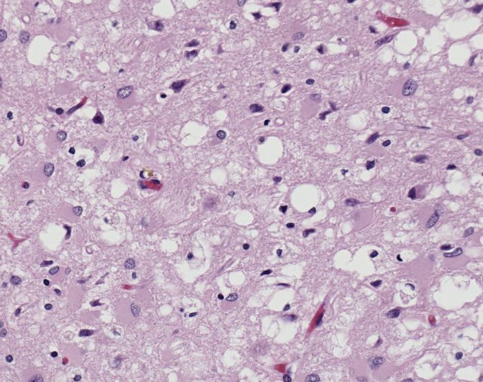
ID#: 10131 Magnified 100X, and stained with H&E (hematoxylin and eosin) staining technique, this light photomicrograph of brain tissue reveals the presence of prominent spongiotic changes in the cortex, and loss of neurons in a case of variant Creutzfeldt-Jakob disease (vCJD). Variant Creutzfeldt-Jakob disease (vCJD) is a prion disease that was first described in 1996 in the United Kingdom. There is now strong scientific evidence that the agent responsible for the outbreak of prion disease in cows, bovine spongiform encephalopathy (BSE or 'mad cow' disease), is the same agent responsible for the outbreak of vCJD in humans. Both disorders are invariably fatal brain diseases with unusually long incubation periods measured in years, and are caused by an unconventional transmissible agent called a prion. vCJD is not the same disease as classic CJD. It has different clinical and pathologic characteristics from classic CJD. Each disease also has a particular genetic profile of the prion protein gene. |
| 原典 | Public Health Image Library (PHIL) ID#: 10131 |
| 作者 |
Content Providers(s): CDC/ Teresa Hammett Photo Credit: Sherif Zaki; MD; PhD; Wun-Ju Shieh; MD; PhD; MPH |
| 許可 (ファイルの再利用) |
Copyright Restrictions: None - This image is in the public domain and thus free of any copyright restrictions. As a matter of courtesy we request that the content provider be credited and notified in any public or private usage of this image. |
ライセンス
この画像は、アメリカ合衆国保健福祉省の一部である疾病予防管理センターの著作物であり、職員の公務の一環として撮影または作成されたものです。アメリカ合衆国連邦政府の著作物として、画像はパブリックドメインの状態にあります。
|
ファイルの履歴
過去の版のファイルを表示するには、その版の日時をクリックしてください。
| 日付と時刻 | サムネイル | 寸法 | 利用者 | コメント | |
|---|---|---|---|---|---|
| 現在の版 | 2008年1月30日 (水) 19:55 | | 700 × 554 (80キロバイト) | Patho | {{Information| |Description=ID#: 10131 Magnified 100X, and stained with H&E (hematoxylin and eosin) staining technique, this light photomicrograph of brain tissue reveals the presence of prominent spongiotic changes in the cortex, and loss of neurons in |
ファイルの使用状況
このファイルを使用しているページはありません。
グローバルなファイル使用状況
以下に挙げる他のウィキがこの画像を使っています:
- ar.wikipedia.org での使用状況
- de.wikibooks.org での使用状況
- en.wikipedia.org での使用状況
- es.wikipedia.org での使用状況
- sr.wikipedia.org での使用状況
,_H%26E.jpg)