
ファイル:Histology bse.jpg
高解像度版はありません。
Histology_bse.jpg (700 × 558 ピクセル、ファイルサイズ: 73キロバイト、MIME タイプ: image/jpeg)
ウィキメディア・コモンズのファイルページにある説明を、以下に表示します。
|
| 解説 |
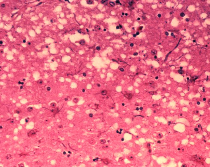
English: This micrograph of brain tissue reveals the cytoarchitectural histopathologic changes found in bovine spongiform encephalopathy. The presence of vacuoles, i.e. microscopic “holes” in the gray matter, gives the brain of BSE-affected cows a sponge-like appearance when tissue sections are examined in the lab.
Nederlands: Deze microscopische opname toont hersenweefsel van een koe die aan BSE gestorven is. Tussen de hersencellen ziet men duidelijk verschillende vacuoles, die deze coupe (weefselsnede) een sponsachtig aanzicht geven.
Deutsch: Das Bild zeigt die histopathologischen Veränderungen die bei einer Infektion mit BSE auftreten. Die Vakuolen, die in der grauen Substanz (substantia grisea) auftreten geben dem Bild ein schwamm-artiges Aussehen.
Français : Cette coupe de tissu cérébral montre les modifications histopathologiques de l'organisation cellulaire lors d'une encéphalopathie spongiforme bovine. la présence de vacuoles, c'est-à-dire des "trous" microscopiques dans le tissu cérébral, donne au cerveau de vaches atteintes de l'ESB un aspect en éponge à l'examen des tissus en laboratoire. |
| 日付 | |
| 原典 | Public Health Image Library, APHIS: http://www.aphis.usda.gov/lpa/issues/bse/bse_photogallery.html |
| 作者 | Dr. Al Jenny |
| その他のバージョン | http://en.wikipedia.org/wiki/Image:Aphis.usda.gov_BSE_5.jpg |
|
|
 |
ファイルの履歴
過去の版のファイルを表示するには、その版の日時をクリックしてください。
| 日付と時刻 | サムネイル | 寸法 | 利用者 | コメント | |
|---|---|---|---|---|---|
| 現在の版 | 2005年6月21日 (火) 10:21 | | 700 × 558 (73キロバイト) | Obarskyr | {{PD}} |
ファイルの使用状況
グローバルなファイル使用状況
以下に挙げる他のウィキがこの画像を使っています:
- af.wikipedia.org での使用状況
- ast.wikipedia.org での使用状況
- as.wikipedia.org での使用状況
- bn.wikipedia.org での使用状況
- bs.wikipedia.org での使用状況
- cs.wikipedia.org での使用状況
- de.wikipedia.org での使用状況
- de.wikibooks.org での使用状況
- el.wikipedia.org での使用状況
- en.wikipedia.org での使用状況
- en.wikibooks.org での使用状況
- en.wikinews.org での使用状況
- eo.wikipedia.org での使用状況
- es.wikipedia.org での使用状況
- eu.wikipedia.org での使用状況
- fa.wikipedia.org での使用状況
- fi.wiktionary.org での使用状況
- fr.wikipedia.org での使用状況
- gl.wikipedia.org での使用状況
- he.wikipedia.org での使用状況
- ht.wikipedia.org での使用状況
- hy.wikipedia.org での使用状況
- ia.wikipedia.org での使用状況
- id.wikipedia.org での使用状況
- is.wikipedia.org での使用状況
- it.wikipedia.org での使用状況
- jv.wikipedia.org での使用状況
- ko.wikipedia.org での使用状況
- lt.wikipedia.org での使用状況
- ms.wikipedia.org での使用状況
- nl.wikipedia.org での使用状況
- no.wikipedia.org での使用状況
- oc.wikipedia.org での使用状況
- pl.wikipedia.org での使用状況
- pnb.wikipedia.org での使用状況
- ro.wikipedia.org での使用状況
このファイルのグローバル使用状況を表示する。