
ファイル:Kinetoplast of Trypanosoma brucei.tif

高解像度版はありません。
Kinetoplast_of_Trypanosoma_brucei.tif (758 × 369 ピクセル、ファイルサイズ: 455キロバイト、MIME タイプ: image/tiff)
ウィキメディア・コモンズのファイルページにある説明を、以下に表示します。
|
概要
| 解説 |
English: Trypanosoma brucei, Electron micrograph of centrin-depleted cells. (a) A typical control cell in this particular section shows single flagellum, pair of basal bodies, kinetoplast and nucleus. (b) TbCen2-depleted cells with multi basal bodies and the kinetoplast with enlarged size compared to the control in ‘a’. Scale bars, 500 nm. F, flagellum; B, basal body; K, kinetoplast; N, nucleus; P, flagellar pocket. |
| 日付 | |
| 原典 | Fig. 3 of: Role of Centrins 2 and 3 in Organelle Segregation and Cytokinesis in Trypanosoma brucei. In: PLOS ONE; doi:10.1371/journal.pone.0045288 |
| 作者 | Angamuthu Selvapandiyan, Praveen Kumar, Jeffrey L. Salisbury, Ching C. Wang, Hira L. Nakhasi |
ライセンス
このファイルはクリエイティブ・コモンズ 表示 2.5 一般ライセンスのもとに利用を許諾されています。
- あなたは以下の条件に従う場合に限り、自由に
- 共有 – 本作品を複製、頒布、展示、実演できます。
- 再構成 – 二次的著作物を作成できます。
- あなたの従うべき条件は以下の通りです。
- 表示 – あなたは適切なクレジットを表示し、ライセンスへのリンクを提供し、変更があったらその旨を示さなければなりません。これらは合理的であればどのような方法で行っても構いませんが、許諾者があなたやあなたの利用行為を支持していると示唆するような方法は除きます。
ファイルの履歴
過去の版のファイルを表示するには、その版の日時をクリックしてください。
| 日付と時刻 | サムネイル | 寸法 | 利用者 | コメント | |
|---|---|---|---|---|---|
| 現在の版 | 2015年1月22日 (木) 03:34 | 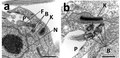 | 758 × 369 (455キロバイト) | Chhandama | User created page with UploadWizard |
ファイルの使用状況
以下のページがこのファイルを使用しています:
グローバルなファイル使用状況
以下に挙げる他のウィキがこの画像を使っています:
- ar.wikipedia.org での使用状況
- bg.wikipedia.org での使用状況
- bs.wikipedia.org での使用状況
- de.wikipedia.org での使用状況
- en.wikipedia.org での使用状況
- it.wikipedia.org での使用状況
- nl.wikipedia.org での使用状況
- ru.wikipedia.org での使用状況