
ファイル:Viral luciferase expression in a mouse tumour.jpg
高解像度版はありません。
Viral_luciferase_expression_in_a_mouse_tumour.jpg (229 × 346 ピクセル、ファイルサイズ: 54キロバイト、MIME タイプ: image/jpeg)
ウィキメディア・コモンズのファイルページにある説明を、以下に表示します。
|
概要
| 解説 |
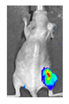
English: A 'nude' (hairless) mouse bearing a tumour in the right hind leg has been treated intra-tumourally with vaccinia virus encoding luciferase. Viral infection of the tumour is visible by the bio-luminescence produced by luciferase. (False coloring is used to demonstrate intensity). Citation: Haddad D, Chen C-H, Carlin S, Silberhumer G, Chen NG, et al. (2012) Imaging Characteristics, Tissue Distribution, and Spread of a Novel Oncolytic Vaccinia Virus Carrying the Human Sodium Iodide Symporter. PLoS ONE 7(8): e41647. doi:10.1371/journal.pone.0041647 |
| 日付 | |
| 原典 | http://www.plosone.org/article/info%3Adoi%2F10.1371%2Fjournal.pone.0041647 |
| 作者 | Haddad D, Chen C-H, Carlin S, Silberhumer G, Chen NG, et al. |
| 許可 (ファイルの再利用) |
http://www.plosone.org/static/license;jsessionid=D9B1E557E36EDD0EDB521F6783647855 |
このファイルはクリエイティブ・コモンズ 表示 1.0 一般ライセンスのもとに利用を許諾されています。
- あなたは以下の条件に従う場合に限り、自由に
- 共有 – 本作品を複製、頒布、展示、実演できます。
- 再構成 – 二次的著作物を作成できます。
- あなたの従うべき条件は以下の通りです。
- 表示 – あなたは適切なクレジットを表示し、ライセンスへのリンクを提供し、変更があったらその旨を示さなければなりません。これらは合理的であればどのような方法で行っても構いませんが、許諾者があなたやあなたの利用行為を支持していると示唆するような方法は除きます。
ライセンス
このファイルはクリエイティブ・コモンズ 表示 2.5 一般ライセンスのもとに利用を許諾されています。
- あなたは以下の条件に従う場合に限り、自由に
- 共有 – 本作品を複製、頒布、展示、実演できます。
- 再構成 – 二次的著作物を作成できます。
- あなたの従うべき条件は以下の通りです。
- 表示 – あなたは適切なクレジットを表示し、ライセンスへのリンクを提供し、変更があったらその旨を示さなければなりません。これらは合理的であればどのような方法で行っても構いませんが、許諾者があなたやあなたの利用行為を支持していると示唆するような方法は除きます。
ファイルの履歴
過去の版のファイルを表示するには、その版の日時をクリックしてください。
| 日付と時刻 | サムネイル | 寸法 | 利用者 | コメント | |
|---|---|---|---|---|---|
| 現在の版 | 2013年5月30日 (木) 17:43 | | 229 × 346 (54キロバイト) | Viraltonic | {{subst:Upload marker added by en.wp UW}} {{Information |Description = {{en|A 'nude' (hairless) mouse bearing a tumour in the right hind leg has been treated intra-tumourally with vaccinia virus encoding luciferase. Viral infection of the tumour is vis... |
ファイルの使用状況
以下のページがこのファイルを使用しています:
グローバルなファイル使用状況
以下に挙げる他のウィキがこの画像を使っています:
- ar.wikipedia.org での使用状況
- en.wikipedia.org での使用状況
- sv.wikipedia.org での使用状況