ファイル:Hippel Lindau.gif

ページのコンテンツが他言語でサポートされていません。

元のファイル(805 × 949 ピクセル、ファイルサイズ: 278キロバイト、MIME タイプ: image/gif)

概要

解説
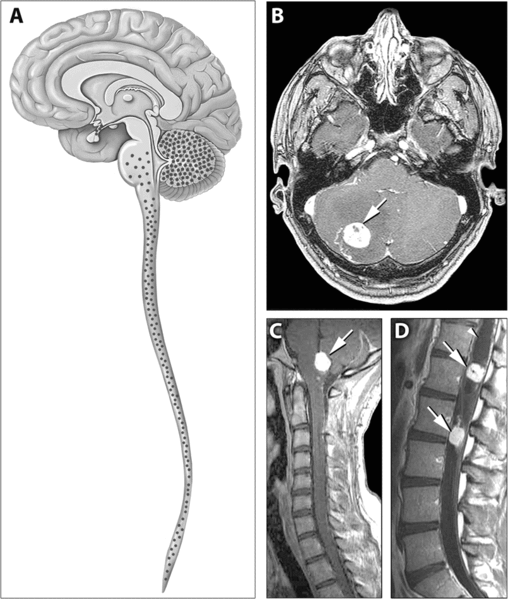
English: Distribution of Hemangioblastomas in the Central Nervous Systems of Study Patients

(A) Schematic representation of the distribution of CNS hemangioblastomas (red dots) in the 25 von Hippel-Lindau disease patients on MRI. Most (98%) of hemangioblastomas were found below the level of the tentorium in the cerebellum, brainstem, and spinal cord.

(B–D) Contrast-enhanced MRI demonstrating representative locations of hemangioblastomas including the cerebellum (B), brainstem (C) and spinal cord (D). (B) Axial view through the cerebellum demonstrating a hyperintense enhancing hemangioblastoma (arrow) with surrounding edema (hypointense area surrounding the tumor) that frequently is associated with these lesions. (C) Sagittal view through the posterior fossa demonstrating a hyperintense enhancing brainstem (medullary) hemangioblastoma (arrow) with surrounding edema. (D) Sagittal view through the thoracic and lumbar spinal cord demonstrating two hyperintense enhancing hemangioblastomas (arrows). The superior tumor is associated with a large intraspinal cyst (syrinx) that is common with these neoplasms (arrowhead)
原典 http://medicine.plosjournals.org/perlserv/?request=get-document&doi=10.1371/journal.pmed.0040060
作者

ライセンス

w:ja:クリエイティブ・コモンズ
表示
このファイルはクリエイティブ・コモンズ 表示 2.5 一般ライセンスのもとに利用を許諾されています。
あなたは以下の条件に従う場合に限り、自由に
  • 共有 – 本作品を複製、頒布、展示、実演できます。
  • 再構成 – 二次的著作物を作成できます。
あなたの従うべき条件は以下の通りです。
  • 表示 – あなたは適切なクレジットを表示し、ライセンスへのリンクを提供し、変更があったらその旨を示さなければなりません。これらは合理的であればどのような方法で行っても構いませんが、許諾者があなたやあなたの利用行為を支持していると示唆するような方法は除きます。
PLOS
PLOS
この画像はオープンアクセスの学術雑誌Public Library of Scienceで公表されたものです。PLoSはサイト上[1]において、刊行する全ての雑誌のコンテンツを、クリエイティブ・コモンズ 表示-2.5 の条件の元で公開する事を宣言しています[2]

アップロードされた方へ: ファイルまたは元論文へのURLリンクを記入しておく必要があります。

Category:Media from PLOS journals配下の適切なカテゴリを付加してください。

キャプション

このファイルの内容を1行で記述してください

このファイルに描写されている項目

題材

ファイルの履歴

過去の版のファイルを表示するには、その版の日時をクリックしてください。

日付と時刻サムネイル寸法利用者コメント
現在の版2007年5月31日 (木) 13:432007年5月31日 (木) 13:43時点における版のサムネイル805 × 949 (278キロバイト)Filip emDistribution of Hemangioblastomas in the Central Nervous Systems of Study Patients (A) Schematic representation of the distribution of CNS hemangioblastomas (red dots) in the 25 von Hippel-Lindau disease patients on MRI. Most (98%) of hemangioblastomas w

以下のページがこのファイルを使用しています:

グローバルなファイル使用状況

以下に挙げる他のウィキがこの画像を使っています: