
ファイル:Determinants of Gastric Acid Secretion.svg

この SVG ファイルのこの PNG プレビューのサイズ: 800 × 550 ピクセル. その他の解像度: 320 × 220 ピクセル | 640 × 440 ピクセル | 1,024 × 704 ピクセル | 1,280 × 880 ピクセル | 2,560 × 1,760 ピクセル | 1,206 × 829 ピクセル。
元のファイル (SVG ファイル、1,206 × 829 ピクセル、ファイルサイズ: 741キロバイト)
ウィキメディア・コモンズのファイルページにある説明を、以下に表示します。
|
概要
| 解説 |
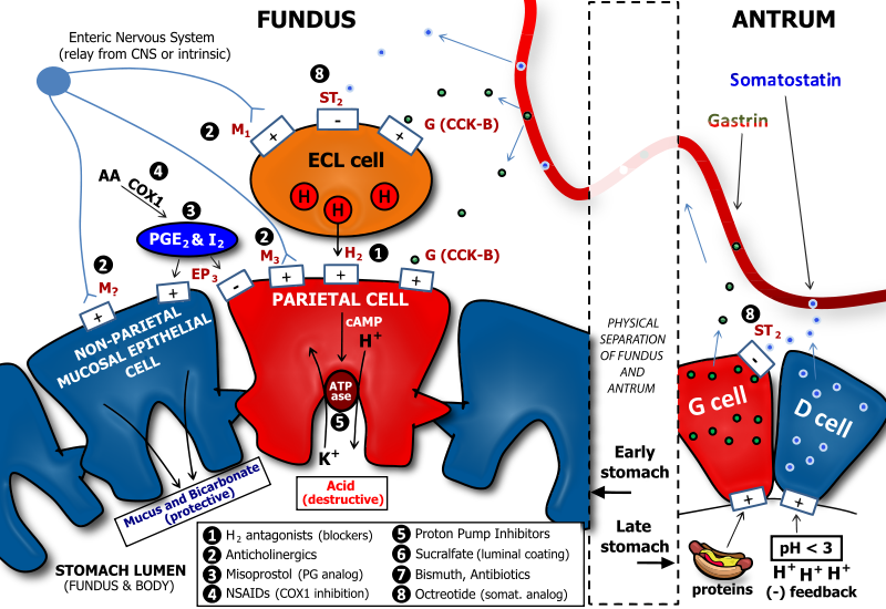
English: There are several neuronal, endocrine, and paracrine determinants of gastric acid secretion. Parietal cells in the fundus and body of the stomach are responsible for secretion of acid into the stomach. Major receptors on parietal cells that trigger acid release when activated include the M3 muscarinic, the H2 histaminic, and the CCK-B or gastrin receptor. The respective ligands for these receptors are acetylcholine, histamine, and gastrin. Acetylcholine is released from enteric neurons, histamine is released from enterochromaffin-like cells, and gastrin is released from G cells which are generally found in the antrum, or lower portion, of the stomach. Enterochromaffin-like (ECL) cells are generally found below the epithelial cell layer of the stomach lumen. ECL cells express M3 muscarinic, ST2 somatostatin, and CCK-B gastrin receptors. While M3 and CCK-B receptors trigger histamine release when activated, the ST2 somatostatin receptor inhibits histamine release. Thus, gastrin is pro-acid, and somatostatin is anti-acid secretion. Both somatostatin and gastrin are released primarily from cells in the antrum. G cells secrete gastrin, and D cells secrete somatostatin. D cells have a regulatory role over G cells in that the somatostatin that they release has an inhibitory influence on gastrin secretion from G cells. This serves to reduce futile simultaneous pro-acid and anti-acid signals to the fundus and body of the stomach. A major trigger for gastrin release is the presence of protein in the antrum: this serves to enhance acid secretion, which is necessary for the activity of pepsin, a major digestive enzyme in the stomach which is important for protein breakdown. In addition, D cells are also subject to regulation by gastric luminal contents. In particular, D cells release more somatostatin when the antral pH is low (i.e., when a highly acidic condition exists). This serves as a negative feedback when acid levels are high, as somatostatin will be secreted, enter the gastric bloodstream, and signal to ECL cells to reduce histamine release. Reduced histamine release will, in turn, reduce acid secretion from parietal cells. Finally, our gastric cells would be susceptible to damage from acid and pepsin if they had no protection. Protection of the gastric mucosal cells is in two forms, a physical barrier of viscous mucus, and a chemical defense of bicarbonate, which neutralizes acid. Protective prostaglandins, i.e., PGE2 and PGI2 activate EP3 receptors on non-parietal mucosal cells to enhance mucus and bicarbonate secretion. At the same time, EP3 receptors are activated on parietal cells to inhibit acid secretion. This is primarily a result of EP3's coupling to the G-protein, Gi, which reduces cyclic adenosine monophosphate (cAMP) levels. The proton pump in parietal cells is an exchanger of potassium and protons. Many portions of the physiology of gastric acid secretion are influenced by drugs, especially those used to treat peptic ulcer disease (PUD) and gastroesophageal reflux disease (GERD). The drug classes and sites of action are denoted by numbers. Note: non-steroidal anti-inflammatory drugs (NSAIDs) such as aspirin and ibuprofen inhibit the production of protective prostaglandins by inhibiting cyclooxygenase 1 (COX1). For this reason, NSAIDs can promote ulcer formation, and their antiplatelet effects also make patients more susceptible to excessive bleeding if a gastric ulcer becomes severe. |
| 日付 | |
| 原典 | 投稿者自身による著作物 |
| 作者 | Adam L. VanWert, Pharm.D., Ph.D. |
| その他のバージョン | このファイルの派生的著作物: Determinants of Gastric Acid Secretion Edit.svg |
ライセンス
この作品の著作権者である私は、この作品を以下のライセンスで提供します。
このファイルはクリエイティブ・コモンズ 表示 3.0 非移植ライセンスのもとに利用を許諾されています。
- あなたは以下の条件に従う場合に限り、自由に
- 共有 – 本作品を複製、頒布、展示、実演できます。
- 再構成 – 二次的著作物を作成できます。
- あなたの従うべき条件は以下の通りです。
- 表示 – あなたは適切なクレジットを表示し、ライセンスへのリンクを提供し、変更があったらその旨を示さなければなりません。これらは合理的であればどのような方法で行っても構いませんが、許諾者があなたやあなたの利用行為を支持していると示唆するような方法は除きます。
ファイルの履歴
過去の版のファイルを表示するには、その版の日時をクリックしてください。
| 日付と時刻 | サムネイル | 寸法 | 利用者 | コメント | |
|---|---|---|---|---|---|
| 現在の版 | 2011年1月16日 (日) 11:03 | | 1,206 × 829 (741キロバイト) | Vanwa71 | .. |
| 2011年1月16日 (日) 10:57 |  | 1,206 × 829 (486キロバイト) | Vanwa71 | Reverted to version as of 10:39, 16 January 2011 | |
| 2011年1月16日 (日) 10:55 |  | 1,206 × 829 (1.33メガバイト) | Vanwa71 | Linked instead of embedded new graphics last time. | |
| 2011年1月16日 (日) 10:39 |  | 1,206 × 829 (486キロバイト) | Vanwa71 | Reverted to version as of 06:03, 16 January 2011 | |
| 2011年1月16日 (日) 10:37 |  | 1,206 × 829 (437キロバイト) | Vanwa71 | cool effects | |
| 2011年1月16日 (日) 06:03 |  | 1,206 × 829 (486キロバイト) | Vanwa71 | Small detail edits. | |
| 2011年1月16日 (日) 05:57 |  | 1,206 × 829 (484キロバイト) | Vanwa71 | Fixed gastrin and somatostatin flaws | |
| 2011年1月16日 (日) 05:14 |  | 1,206 × 829 (472キロバイト) | Vanwa71 | Forgot categories. | |
| 2011年1月16日 (日) 05:11 |  | 1,206 × 829 (472キロバイト) | Vanwa71 | I finally started using my head, and converted all the text to outlines to keep it exactly as I wanted it. | |
| 2011年1月16日 (日) 05:05 |  | 1,206 × 829 (214キロバイト) | Vanwa71 | Please bear with me, I finally looked up websafe fonts instead of trial and error. Let's hope this works. |
ファイルの使用状況
以下のページがこのファイルを使用しています:
グローバルなファイル使用状況
以下に挙げる他のウィキがこの画像を使っています:
- el.wikipedia.org での使用状況
- en.wikipedia.org での使用状況
- fr.wikipedia.org での使用状況
- hi.wikipedia.org での使用状況
- id.wikipedia.org での使用状況
- ko.wikipedia.org での使用状況
- sq.wikipedia.org での使用状況
- te.wikipedia.org での使用状況
- zh.wikipedia.org での使用状況
- zh.wikibooks.org での使用状況