
ファイル:PDA Coil.png
高解像度版はありません。
PDA_Coil.png (636 × 432 ピクセル、ファイルサイズ: 106キロバイト、MIME タイプ: image/png)
ウィキメディア・コモンズのファイルページにある説明を、以下に表示します。
|
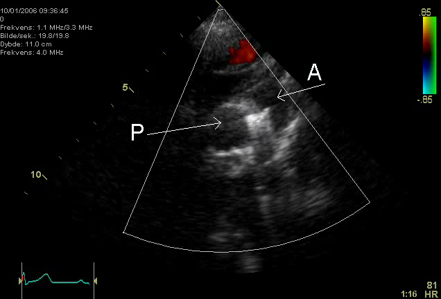
An echocardiogram of a coiled persisting ductus arteriosus. One can see the aortic arch,the pulmonary artery and the coil between them.
Image made by myself, Kjetil Lenes. Released in agreement with my employer.
| この著作物の著作権者である私は、この著作物における権利を放棄しパブリックドメインとします。これは全世界で適用されます。 一部の国では、これが法的に可能ではない場合があります。その場合は、次のように宣言します。 私は、あらゆる人に対して、法により必要とされている条件を除き、如何なる条件も課すことなく、あらゆる目的のためにこの著作物を使用する権利を与えます。 |
Keywords:
- en: Echocardiography, Sonography, Ultrasonography, Medical Ultrasound, Cardiology, persisting ductus arteriosus
- de: Echokardiographie, Sonografie, Sonographie, Ultraschall, Kardiologie, ductus arteriosus
ファイルの履歴
過去の版のファイルを表示するには、その版の日時をクリックしてください。
| 日付と時刻 | サムネイル | 寸法 | 利用者 | コメント | |
|---|---|---|---|---|---|
| 現在の版 | 2006年2月18日 (土) 16:50 | | 636 × 432 (106キロバイト) | Ekko | An echocardiogram of a coiled persisting ductus arteriosus. One can see the aortic arch,the pulmonary artery and the coil between them. Image made by myself, Kjetil Lenes. Released in agreement with my employer. |
ファイルの使用状況
以下のページがこのファイルを使用しています:
グローバルなファイル使用状況
以下に挙げる他のウィキがこの画像を使っています:
- ar.wikipedia.org での使用状況
- en.wikipedia.org での使用状況
- fa.wikipedia.org での使用状況
- he.wikipedia.org での使用状況
- nn.wikipedia.org での使用状況