
ファイル:Microvilli.jpg
高解像度版はありません。
Microvilli.jpg (640 × 448 ピクセル、ファイルサイズ: 83キロバイト、MIME タイプ: image/jpeg)
ウィキメディア・コモンズのファイルページにある説明を、以下に表示します。
|
概要
| 解説 |
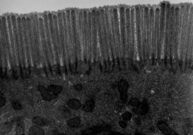
English: Transmission electron microscope image of a thin section cut through a human jejunum (segment of small intestine) epithelial cell. Image shows apical end of absorptive cell with some of the densely packed microvilli that make up the striated border. Each microvillus is approximately 1um long by 0.1um in diameter and contains a core of actin microfilaments. |
| 原典 | http://remf.dartmouth.edu/images/humanMicrovilliTEM/source/1.html |
| 作者 | Louisa Howard, Katherine Connollly - Dartmouth Electron Microscope Facility |
| 許可 (ファイルの再利用) |
http://remf.dartmouth.edu/imagesindex.html |
ライセンス
| この著作物は、著作者であるDartmouth Electron Microscope Facilityによって権利が放棄され、パブリックドメインとされました。これは全世界で適用されます。 一部の国では、これが法的に可能ではない場合があります。その場合は、次のように宣言します。 Dartmouth Electron Microscope Facilityは、あらゆる人に対して、法により必要とされている条件を除き、如何なる条件も課すことなく、あらゆる目的のためにこの著作物を使用する権利を与えます。
|
このファイルは、18:28, 13 October 2011 (UTC)に管理者又は信頼された利用者であるCommon Good (talk)によって査読され、その時点で、パブリックドメインの状態にあることが確認されました。
|
元のアップロードログ
Originally from en.wikipedia; description page is (was) here
- 18:51, 28 December 2002 Magnus Manske 350x247 (19,689 bytes) (Source and public domain notice at [http://remf.dartmouth.edu/imagesindex.html])
ファイルの履歴
過去の版のファイルを表示するには、その版の日時をクリックしてください。
| 日付と時刻 | サムネイル | 寸法 | 利用者 | コメント | |
|---|---|---|---|---|---|
| 現在の版 | 2015年11月17日 (火) 22:00 | | 640 × 448 (83キロバイト) | SteinsplitterBot | Bot: Image rotated by 180° |
| 2011年10月13日 (木) 18:23 |  | 640 × 455 (82キロバイト) | Common Good | high res | |
| 2006年5月9日 (火) 19:42 |  | 350 × 247 (19キロバイト) | Magnus Manske | {{Information| |Description= Source and public domain notice at [http://remf.dartmouth.edu/imagesindex.html] * 18:51, 28 December 2002 Magnus Manske 350x247 (19,689 bytes) ''<nowiki>(Source and public domain notice at [http://r |
ファイルの使用状況
以下のページがこのファイルを使用しています:
グローバルなファイル使用状況
以下に挙げる他のウィキがこの画像を使っています:
- az.wikipedia.org での使用状況
- bn.wikipedia.org での使用状況
- cy.wikipedia.org での使用状況
- de.wiktionary.org での使用状況
- dv.wikipedia.org での使用状況
- en.wikipedia.org での使用状況
- en.wikiversity.org での使用状況
- es.wikipedia.org での使用状況
- fa.wikipedia.org での使用状況
- fr.wikipedia.org での使用状況
- gl.wikipedia.org での使用状況
- hu.wikipedia.org での使用状況
- hy.wikipedia.org での使用状況
- it.wikipedia.org での使用状況
- ko.wikipedia.org での使用状況
- lt.wikipedia.org での使用状況
- pam.wikipedia.org での使用状況
- tr.wikipedia.org での使用状況