
ファイル:Macs killing cancer cell.jpg

このプレビューのサイズ: 800 × 583 ピクセル。 その他の解像度: 320 × 233 ピクセル | 640 × 467 ピクセル | 1,024 × 747 ピクセル | 1,280 × 933 ピクセル | 2,289 × 1,669 ピクセル。
元のファイル (2,289 × 1,669 ピクセル、ファイルサイズ: 1.08メガバイト、MIME タイプ: image/jpeg)
ウィキメディア・コモンズのファイルページにある説明を、以下に表示します。
|
概要
| 解説 |
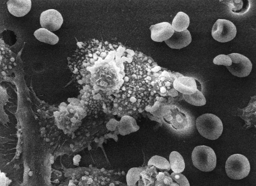
English: [Part of a] six-step sequence of the death of a cancer cell. A cancer cell has migrated through the holes of a matrix coated membrane from the top to the bottom, simulating natural migration of a invading cancer cell between, and sometimes through, the vascular endothelium. Notice the spikes or pseudopodia that are characteristic of an invading cancer cell (1). A buffy coat containing red blood cells, lymphocytes and macrophages is added to the bottom of the membrane. A group of macrophages identify the cancer cell as foreign matter and start to stick to the cancer cell, which still has its spikes (2). Shown: Macrophages begin to fuse with, and inject its toxins into, the cancer cell. The cell starts rounding up and loses its spikes (3). As the macrophage cell becomes smooth (4). The cancer cell appears lumpy in the last stage before it dies. These lumps are actually the macrophages fused within the cancer cell (5). The cancer cell then loses its morphology, shrinks up and dies (6). Photo magnification: 3: x8,000 Type: B & W print العربية : سلسلة من ست خطوات لموت خلية سرطانية. هاجرت خلية سرطانية عبر فتحات من الغشاء المكسو بالمطرس من الأعلى إلى الأسفل محاكية الهجرة الطبيعية للخلية السرطانية الغازية بين -وأحيانا عبر- البطانة الوعائية. لاحظ الشوكات أو الأقدام الكاذبة المميِّزة للخلية السرطانية الغازية (1). يُضاف كساء (طبقة) تحتوي على خلايا الدم الحمراء واللمفاويات والبالعات الكبيرة إلى أسفل الغشاء. تتعرف البالعات الكبيرة على الخلية السرطانية على أنها مادة دخيلة وتبدأ بالالتصاق بها وهي مازالت تملك شوكاتها (2). ظاهر في الصورة: تبدأ البالعات الكبيرة في الاندماج وحقن السموم داخل الخلية السرطانية. تبدأ الخلية السرطانية في اتخاذ شكل دائري وتفقد شوكاتها (3). بينما تصبح البالعة الكبيرة ملساء (4). تظهر الخلايا السرطانية متكتلة ومتنتئة في المرحلة الأخيرة قبل موتها. الكتل والنتوءات هي بالعات كبيرة مندمجة داخل الخلية السرطانية (5). تفقد الخلية السرطانية شكلها بعد ذلك وتتقلص ثم تموت (6). تكبير الصورة: 3: x8,000، ونوعها: نسخة بالأبيض والأسود. |
||||||
| 日付 | Date Created: October 1988 | ||||||
| 原典 | Image and description: Dr. Raowf Guirguis. National Cancer Institute | ||||||
| 作者 | Susan Arnold (photographer) | ||||||
| 許可 (ファイルの再利用) |
|
||||||

ファイルの履歴
過去の版のファイルを表示するには、その版の日時をクリックしてください。
| 日付と時刻 | サムネイル | 寸法 | 利用者 | コメント | |
|---|---|---|---|---|---|
| 現在の版 | 2006年10月4日 (水) 03:16 | | 2,289 × 1,669 (1.08メガバイト) | DO11.10 | |
| 2006年10月4日 (水) 03:15 |  | 2,289 × 1,800 (1.02メガバイト) | DO11.10 | {{Information |Description=[Part of a] six-step sequence of the death of a cancer cell. A cancer cell has migrated through the holes of a matrix coated membrane from the top to the bottom, simulating natural migration of a invading cancer cell between, an |
ファイルの使用状況
以下のページがこのファイルを使用しています:
グローバルなファイル使用状況
以下に挙げる他のウィキがこの画像を使っています:
- ar.wikipedia.org での使用状況
- ast.wikipedia.org での使用状況
- az.wikipedia.org での使用状況
- bg.wikipedia.org での使用状況
- ca.wikipedia.org での使用状況
- cs.wikipedia.org での使用状況
- de.wikibooks.org での使用状況
- en.wikipedia.org での使用状況
- es.wikipedia.org での使用状況
- et.wikipedia.org での使用状況
- eu.wikipedia.org での使用状況
- fa.wikipedia.org での使用状況
- gl.wikipedia.org での使用状況
- he.wikipedia.org での使用状況
- hu.wikipedia.org での使用状況
- id.wikipedia.org での使用状況
- it.wikipedia.org での使用状況
- nds.wikipedia.org での使用状況
- nl.wikipedia.org での使用状況
- pt.wikipedia.org での使用状況
- pt.wikiversity.org での使用状況
- sl.wikipedia.org での使用状況
- sq.wikipedia.org での使用状況
- tr.wikipedia.org での使用状況
- uz.wikipedia.org での使用状況
- vi.wikipedia.org での使用状況
- www.wikidata.org での使用状況
- zh.wikipedia.org での使用状況